File:Proximal fractures of 5th metatarsal.jpg
Jump to navigation
Jump to search

Size of this preview: 578 × 599 pixels. Other resolutions: 231 × 240 pixels | 463 × 480 pixels | 579 × 600 pixels | 741 × 768 pixels | 988 × 1,024 pixels | 1,709 × 1,772 pixels.
Original file (1,709 × 1,772 pixels, file size: 666 KB, MIME type: image/jpeg)
Summary
| Description |
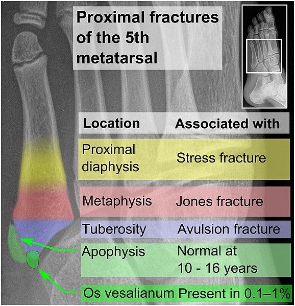
English: Proximal fractures of 5th metatarsal: Proximal fractures of the fifth metatarsal are common,[1] and are distinguished by their locations:
Normal anatomy that may simulate a fracture include mainly:
Template in WikipediaTo edit image template in Wikipedia, go to: en:Template:Image of proximal fractures of the 5th metatarsal. Further reading |
|||
| Date | ||||
| Source |
References
|
|||
| Author |
.jpg) - Reusing images - Conflicts of interest: None |
|||
| Other versions |
|


Licensing
| This file is made available under the Creative Commons CC0 1.0 Universal Public Domain Dedication. | |
| The person who associated a work with this deed has dedicated the work to the public domain by waiving all of their rights to the work worldwide under copyright law, including all related and neighboring rights, to the extent allowed by law. You can copy, modify, distribute and perform the work, even for commercial purposes, all without asking permission.
|
File history
Click on a date/time to view the file as it appeared at that time.
| Date/Time | Thumbnail | Dimensions | User | Comment | |
|---|---|---|---|---|---|
| current | 11:06, 29 July 2019 | | 1,709 × 1,772 (666 KB) | Mikael Häggström | Singular |
File usage
The following page uses this file: