Uncategorized files
Jump to navigation
Jump to search
Showing below up to 50 results in range #1 to #50.
View (previous 50 | next 50) (20 | 50 | 100 | 250 | 500)
 ?don't recall.png 500 × 500; 182 KB
 Abdomen and pelvis for menu.png 330 × 659; 387 KB
 Anatomy image for main menu.png 337 × 1,540; 650 KB
 Boy-3402783 960 720.png 894 × 720; 171 KB
 Boy-3402794 960 720.png 832 × 720; 163 KB
 Boy-3402795 960 720.png 960 × 692; 193 KB
 CT of the heart.jpg 509 × 547; 48 KB
 CT of the liver.jpg 216 × 214; 22 KB
 Causes of hip pain.jpg 458 × 218; 36 KB
 Contrast-enhanced ultrasonography of benign lesion - local.jpg 956 × 503; 116 KB
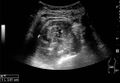 Cortical solid mass of renal cell carcinoma - local.jpg 1,012 × 698; 106 KB
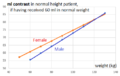 Diagram - weight.png 476 × 300; 13 KB
 Logo.png 135 × 135; 11 KB
 Look of Radlines on the day of its creation.png 1,076 × 966; 158 KB
 Measures of the kidney - local.jpg 1,024 × 1,659; 258 KB
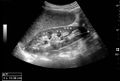 Normal adult kidney - local.jpg 1,007 × 681; 106 KB
 Normal pediatric kidney - local.jpg 1,011 × 634; 105 KB
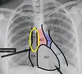 Optimal central venous catheter tip location.jpg 443 × 402; 52 KB
 Patholines logo.png 135 × 135; 7 KB
 Radviser.logo.png 135 × 135; 12 KB
 Reimer's migration index (crop).png 726 × 345; 252 KB
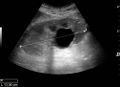 Simple cyst with posterior enhancement - local.jpg 1,008 × 728; 101 KB
 Spondylolisthesis - crop.jpg 161 × 158; 7 KB
 Spondylolisthesis - crop X-ray.jpg 151 × 164; 5 KB
 Strain elastography of a normal kidney - local.jpg 1,016 × 563; 152 KB
 Ultrasonography of acute pyelonephritis - local.jpg 523 × 390; 45 KB
 Ultrasonography of angiomyolipoma - local.jpg 1,010 × 812; 137 KB
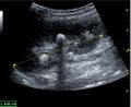 Ultrasonography of centrally-located stone with posterior shadowing - local.jpg 1,009 × 820; 137 KB
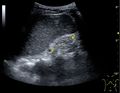 Ultrasonography of end-stage chronic kidney disease - local.jpg 962 × 743; 110 KB
 Ultrasonography of kidney with nephrotic syndrome - local.jpg 747 × 620; 69 KB
 Ultrasonography of left hydroureter with ureteric jet - local.jpg 990 × 606; 99 KB
 Ultrasonography of multiple angiomyolipomas in tuberous sclerosis - local.jpg 1,007 × 703; 125 KB
 Ultrasonography of percutaneous nephrostomy tube - local.jpg 773 × 384; 69 KB
 Ultrasonography of postoperative renal failure - local.jpg 688 × 573; 87 KB
 Ultrasonography of renal lymphoma - local.jpg 1,008 × 968; 139 KB
 Ultrasonography of renal trauma with laceration - local.jpg 598 × 500; 66 KB


,_diastolic_velocity_(Vd),_acceleration_time_(AoAT),_systolic_acceleration_(Ao_Accel)_and_resistive_index_(RI)_of_normal_kidney_-_local.jpg)


.png)


