Template:Hip dysplasia - choice of modality
Author:
Mikael Häggström [notes 1]
Choice of modality
| Age | Scenario | Usual appropriate initial imaging |
|---|---|---|
| <4 weeks | Equivocal physical examination or risk factors | No imaging |
| Physical findings of DDH | Ultrasonography | |
| 4 weeks - 4 months | Equivocal physical examination or risk factors | Ultrasonography |
| 4 - 6 months | Concern for DDH | X-ray. Ultrasonography may be appropriate[notes 2] |
| >6 months | X-ray |
Hip dysplasia diagnosed by ultrasound[2] or projectional radiography (see X-ray of hip dysplasia.[3] Ultrasound imaging is generally preferred at up to 4 months due to limited ossification of the skeleton.[1][notes 2]
- Projectional radiography
- Main article: X-ray of hip dysplasia
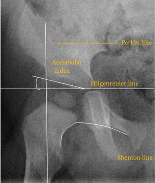
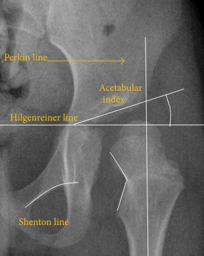
Despite the widespread of ultrasound, pelvis X-ray is still frequently used to diagnose and/or monitor hip dysplasia or for assessing other congenital conditions or bone tumors.[4]
The most useful lines and angles that can be drawn in the pediatric pelvis assessing hip dysplasia are as follows:[4]
Different measurements are used in adults.[4]==Notes==
- ↑ For a full list of contributors, see article history. Creators of images are attributed at the image description pages, seen by clicking on the images. See Radlines:Authorship for details.
- ↑ 2.0 2.1 Ultrasonography is the imaging method of choice up to 6 months for the nonoperative surveillance imaging in harness of known diagnosis of DDH.
- . ACR Appropriateness Criteria - Developmental Dysplasia of the Hip (DDH)–Child. American College of Radiology. Revised 2018
References
- ↑ 1.0 1.1 . ACR Appropriateness Criteria - Developmental Dysplasia of the Hip (DDH)–Child. American College of Radiology. Revised 2018
- ↑ . Ultrasound Detection of DDH - International Hip Dysplasia Institute.
- ↑ . X-Ray Screening for Developmental Dysplasia of the Hip - International Hip Dysplasia Institute.
- ↑ 4.0 4.1 4.2 4.3 4.4 Initially largely copied from: Ruiz Santiago, Fernando; Santiago Chinchilla, Alicia; Ansari, Afshin; Guzmán Álvarez, Luis; Castellano García, Maria del Mar; Martínez Martínez, Alberto; Tercedor Sánchez, Juan (2016). "Imaging of Hip Pain: From Radiography to Cross-Sectional Imaging Techniques ". Radiology Research and Practice 2016: 1–15. doi:. ISSN 2090-1941. Attribution 4.0 International (CC BY 4.0) license

