Ultrasonography of cirrhosis
Author:
Mikael Häggström [notes 1]
Planning
- Need of investigation
The diagnosis of cirrhosis can be made by physical examination and blood testing alone,[1] but ultrasonography is very helpful as an additional diagnostic tool at least in subtle disease.
- Choice of modality
Ultrasonography is the first-line modality of medical imaging in patients with suspected cirrhosis and/or portal hypertension.[2] US is also typically the initial, first-line modality choice for the diagnosis and follow-up of portal hypertension.[3] For cirrhosis, it should be done without IV contrast.
How soon
In Swedish practice, ultrasonography for suspected cirrhosis should be done within 2 months.[notes 2]
In patients with known cirrhosis, surveillance ultrasonography of hepatocellular cancer (HCC) is indicated, generally every 6 months.[4]
Diagnosis
Cirrhosis is indicated by:
- Surface nodularity: (sensitivity 88%, specificity 82-95%)[5]
- General coarse and heterogeneous texture[5]
- Segmental hypertrophy or atrophy[5]
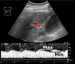
- Portal vein measurement and Doppler:
- Hepatic artery Doppler:
- Splenomegaly[5] (see ultrasonography of the spleen for size cutoffs)
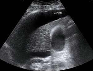
- Ascites,[5] with fluid seen in the hepatorenal recess.
- For further signs of portal hypertension, which may be used upon specific portal hypertension referral, see Ultrasonography of portal hypertension
- Portal flow pulsatility: An increased portal vein pulsatility is an indicator of cirrhosis (even without portal hypertension), but may also be caused by an increased right atrial pressure.[6] Portal vein pulsatility can be quantified by pulsatility indices (PI), where an index above a certain cutoff indicates pathology:
| Index | Calculation | Cutoff |
|---|---|---|
| Average-based | (Max - Min) / Average[6] | 0.5[6] |
| Max-relative | (Max - Min) / Max[7] | 0.5[7][8] - 0.54[8] |
Surveillance
In patients with a diagnosis of cirrhosis, surveillance ultrasonography of hepatocellular cancer (HCC) is indicated, generally every 6 months.[4]
Notes
- ↑ For a full list of contributors, see article history. Creators of images are attributed at the image description pages, seen by clicking on the images. See Radlines:Authorship for details.
- ↑ NU Hospital Group, Sweden
References
- ↑ Udell, JA; Wang, CS; Tinmouth, J; FitzGerald, JM; Ayas, NT; Simel, DL; Schulzer, M; Mak, E; et al. (Feb 22, 2012). "Does this patient with liver disease have cirrhosis? ". JAMA: The Journal of the American Medical Association 307 (8): 832–42. doi:. PMID 22357834.
- ↑ Procopet, Bogdan; Berzigotti, Annalisa (2017). "Diagnosis of cirrhosis and portal hypertension: imaging, non-invasive markers of fibrosis and liver biopsy ". Gastroenterology Report 5 (2): 79–89. doi:. ISSN 2052-0034.
- ↑ Bandali, Murad Feroz; Mirakhur, Anirudh; Lee, Edward Wolfgang; Ferris, Mollie Clarke; Sadler, David James; Gray, Robin Ritchie; Wong, Jason Kam (2017). "Portal hypertension: Imaging of portosystemic collateral pathways and associated image-guided therapy ". World Journal of Gastroenterology 23 (10): 1735. doi:. ISSN 1007-9327.
- ↑ 4.0 4.1 Fateen, Waleed; Ryder, Stephen (2017). "Screening for hepatocellular carcinoma: patient selection and perspectives ". Journal of Hepatocellular Carcinoma Volume 4: 71–79. doi:. ISSN 2253-5969.
- ↑ 5.00 5.01 5.02 5.03 5.04 5.05 5.06 5.07 5.08 5.09 5.10 5.11 5.12 5.13 5.14 Dr Yuranga Weerakkody, A.Prof Frank Gaillard et al.. Cirrhosis. Radiopaedia. Retrieved on 2018-06-16.
- ↑ 6.0 6.1 6.2 Iranpour, Pooya; Lall, Chandana; Houshyar, Roozbeh; Helmy, Mohammad; Yang, Albert; Choi, Joon-Il; Ward, Garrett; Goodwin, Scott C (2016). "Altered Doppler flow patterns in cirrhosis patients: an overview ". Ultrasonography 35 (1): 3–12. doi:. ISSN 2288-5919.
- ↑ 7.0 7.1 Goncalvesova, E.; Varga, I.; Tavacova, M.; Lesny, P. (2013). "Changes of portal vein flow in heart failure patients with liver congestion ". European Heart Journal 34 (suppl 1): P627–P627. doi:. ISSN 0195-668X.
- ↑ 8.0 8.1 Page 367 in: Henryk Dancygier (2009). Clinical Hepatology: Principles and Practice of Hepatobiliary Diseases . 1. Springer Science & Business Media. ISBN 9783540938422.

