Ultrasonography of kidney stone disease
Authors:
Mikael Häggström; Authors of Creative Commons article[1] [notes 1]
This article deals with acute kidney stone disease. For non-emergent exams, see CT in recurrent kidney stone disease.
Contents
Planning
Need for imaging
Imaging is indicated in cases of flank pain and hematuria.[2]
Choice of modality

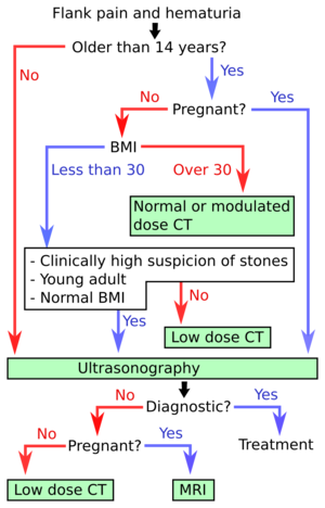
- Ultrasonography of kidney stone disease is the first-line imaging modality for patients <14 years of age and those who are pregnant. It is also the first-line investigation for thin (BMI <30) patients and there is a strong suspicion of kidney stone disease.[2] In the algorithm at right, hydronephrosis may count as a diagnostic finding of urolithiasis.
- CT of kidney stone disease is recommended for older patients, as well as those who have higher BMI and/or less specific findings.[2]
How soon
Within a few hours.
Evaluation
With ultrasonography, larger stones (>5–7 mm) within the kidney, i.e., in the calyces, the pelvis and the pyeloureteric junction, can be differentiated, especially in the cases with accompanying hydronephrosis. Hyperechoic stones are seen with accompanying posterior shadowing. Additional twinkling artifacts below the stone can often be seen using Doppler US.
Renal stone located at the pyeloureteric junction with accompanying hydronephrosis.[1]
Centrally-located stone with posterior shadowing. No hydronephrosis is present. Measurement of kidney length on the US image is illustrated by ‘+’ and a dashed line.[1]

Large stones filling the entire collecting system are called coral stones or staghorn calculi and are easily visualized with US.
Stones in the ureters are usually not visualized with US due to the air-filled intestines obscuring the insonation window. However, ureteral stones near the ostium can be visualized with a scan position over the bladder. An exam of the ureteric orifices and the excretion of urine to the bladder can be performed by inspecting the ureteric jets in the bladder with color Doppler US.
Left hydroureter with ureteric jet. No stone is visible. The red color in the color box represents motion towards the transducer as defined by the color bar.[1]
Report
- Presence or absence of hydronephrosis.
- Location and size of any stones. The absence of visible stones is not necessary to report, since the high risk of missing them may result in a false reassurance.
- See also: General notes on reporting
Notes
- ↑ For a full list of contributors, see article history. Creators of images are attributed at the image description pages, seen by clicking on the images. See Radlines:Authorship for details.
References
- ↑ 1.0 1.1 1.2 1.3 1.4 Content initially copied from: Hansen, Kristoffer; Nielsen, Michael; Ewertsen, Caroline (2015). "Ultrasonography of the Kidney: A Pictorial Review ". Diagnostics 6 (1): 2. doi:. ISSN 2075-4418. (CC-BY 4.0)
- ↑ 2.0 2.1 2.2 Brisbane, Wayne; Bailey, Michael R.; Sorensen, Mathew D. (2016). "An overview of kidney stone imaging techniques ". Nature Reviews Urology 13 (11): 654–662. doi:. ISSN 1759-4812.
- ↑ Brisbane, Wayne; Bailey, Michael R.; Sorensen, Mathew D. (2016). "An overview of kidney stone imaging techniques ". Nature Reviews Urology 13 (11): 654–662. doi:. ISSN 1759-4812.


