Difference between revisions of "Ultrasonography of steatosis"
Jump to navigation
Jump to search
(→Evaluation: Generalized) |
(+Choice of modality) |
||
| Line 4: | Line 4: | ||
}} | }} | ||
==Planning== | ==Planning== | ||
| + | {{Steatosis - choice of modality}} | ||
===How soon=== | ===How soon=== | ||
Within 2 months in Swedish practice<ref group="notes">{{NU Hospital Group}}</ref> | Within 2 months in Swedish practice<ref group="notes">{{NU Hospital Group}}</ref> | ||
Revision as of 17:01, 15 July 2019
Author:
Mikael Häggström [notes 1]
Planning
Choice of modality
- Ultrasonography of steatosis is a good method for screening. Quantification can be done, but it is not as accurate as MRI.
- MRI of liver steatosis is the most accurate method to quantify liver steatosis.
- CT of the liver should not be used to detect or stage liver steatosis. Steatosis on CT scan is displayed as a liver with attenuation lower than usual. CT has high sensitivity for moderate and severe steatosis, but lower for mild steatosis.
How soon
Within 2 months in Swedish practice[notes 2]
Evaluation

Liver echogenicity, as compared to the kidney, where liver hyperechogenicity indicates steatosis.
It is also appropriate to perform a general upper abdominal screening.
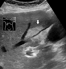
Abdominal ultrasonography of focal steatosis. It can br distinguished from a tumor by absence of signs of expansion, in this case not compressing the hepatic vein going through it.

Report
- Normal: "Normal liver echogenicity."
- Hyperechogenic: "Hyperechogenic liver parenchyma, indicating steatosis".
Notes
- ↑ For a full list of contributors, see article history. Creators of images are attributed at the image description pages, seen by clicking on the images. See Radlines:Authorship for details.
- ↑ NU Hospital Group, Sweden