Ultrasonography of superficial soft tissues
Author:
Mikael Häggström [notes 1]
Contents
Planning
Choice of modality
- Ultrasonography of superficial soft tissues is the investigation of choice in stable superficial soft tissue masses of unknown origin, at least in flexion surfaces of large joints (groin, popliteal fossa, armpit and cubital fossa).
- MRI is the imaging modality of choice in suspected lipoma, with superior sensitivity of distinguishing it from liposarcoma as well as mapping the surrounding anatomy.[1]
Evaluation
Possible findings:
Lymph nodes

A typical normal lymph node: smooth, gently lobulated oval with a hypoechoic cortex measuring less than 3 mm in thickness with a central echogenic hilum.[2]

A suspected malignant lymph node:
- Absence of the fatty hilum
- Increased focal cortical thickness greater than 3 mm
- Doppler ultrasonography that shows hyperaemic blood flow in the hilum and central cortex and/or abnormal (non-hilar cortical) blood flow.[2]


Lipoma/Liposarcoma
A lipoma: hyperechoic compared to adjacent muscle, and relatively well-defined, with miniature hyperechoic lines.[3]

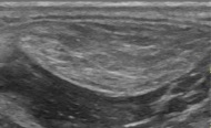
Liposarcoma: In this case a heterogeneous mass consisting of an upper hyperechoic portion, corresponding to lipomatous matrix, and areas of hypoechogenicity corresponding to nonlipomatous components.[4]
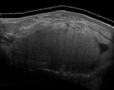
Liposarcoma mimicking lipoma. A homogeneous hypoechoic mass presenting with the same appearance of lipoma. It was clinically distinguished as having rapid growth.[4] It demonstrates the inaccuracy of ultrasound to properly distinguish lipomas from liposarcomas, so this case should have had MRI as a first investigation.


Notes
- ↑ For a full list of contributors, see article history. Creators of images are attributed at the image description pages, seen by clicking on the images. See Radlines:Authorship for details.
References
- ↑ Rohit Sharma and A.Prof Frank Gaillard et al.. Lipoma. Radiopaedia. Retrieved on 2018-09-27.
- ↑ 2.0 2.1 Dialani, V.; James, D. F.; Slanetz, P. J. (2014). "A practical approach to imaging the axilla ". Insights into Imaging 6 (2): 217–229. doi:. ISSN 1869-4101. Creative Commons attribution license
- ↑ Chernev I, Tingey S. (2013). "Thenar intramuscular lipoma: A case report. ". J Med Cases 4 (10): 676-8.
- ↑ 4.0 4.1 Content originally copied from: Mak, Chee-Wai; Tzeng, Wen-Sheng (2012). Sonography of the Scrotum . doi:. from Kerry Thoirs. Sonography. ISBN 978-953-307-947-9Script error: No such module "check isxn"., Published: February 3, 2012, under the CC-BY-3.0 license.