Difference between revisions of "Ultrasonography of the abdomen and pelvis"
(Created) |
m (→General screening: Spaced) |
||
| (34 intermediate revisions by the same user not shown) | |||
| Line 1: | Line 1: | ||
| − | {{ | + | {{Top |
|author1=[[User:Mikael Häggström|Mikael Häggström]] | |author1=[[User:Mikael Häggström|Mikael Häggström]] | ||
|author2= | |author2= | ||
}} | }} | ||
| − | == | + | ==Planning== |
| − | + | ;Choice of exam | |
| − | [[ | + | For patients presenting with symptoms that are less specific for any certain organ, such as unspecific acute [[abdominal pain]], consider an '''[[abdominal/pelvic CT]]''' instead. |
| − | |||
| − | = | + | ;At least 6 hours of fasting<ref group="notes">Otherwise high risk of intestines obscuring the organ of interest.<br>- {{NU Hospital Group}}</ref> |
| − | {{ | + | Exceptions: |
| + | *The target structures are known to be superficial to any intestines | ||
| + | *The case is emergent enough to justify a suboptimal exam | ||
| − | == | + | ==Locations== |
| − | {{ | + | <gallery> |
| + | File:Liver measurements on ultrasonography.jpg|link=Ultrasonography of the liver|[[Ultrasonography of the liver|Ultrasonography of the '''liver''']] | ||
| + | File:Normal adult kidney.jpg|link=Ultrasonography of the urinary system|[[Ultrasonography of the urinary system|Ultrasonography of the '''urinary system''']] | ||
| + | File:Ultrasonography of a normal testicle.jpg|link=Ultrasonography of the scrotum|[[Ultrasonography of the scrotum|Ultrasonography of the '''scrotum''']] | ||
| + | File:Ultrasonography of a normal looking gallbladder.jpg|link=Ultrasonography of the biliary tract|[[Ultrasonography of the biliary tract|Ultrasonography of the '''biliary tract''']] | ||
| + | File:Ultrasonography of diastasis recti.jpg|thumb|[[Ultrasonography of the abdominal wall|Ultrasonography of the '''abdominal wall''']] | ||
| + | </gallery> | ||
| + | |||
| + | ==General screening== | ||
| + | ===Upper abdomen=== | ||
| + | For symptoms of the upper abdomen, it is a proper custom to perform a general screening of the following locations: | ||
| + | *'''Pancreas''', mainly for dilatation of the pancreatic duct, or obvious tumors. | ||
| + | *'''Liver''', for echogenicity and focal changes. <br>''In case of suspected pathology, see: [[Ultrasonography of the liver]]'' | ||
| + | *'''Hepatorenal recess''' for ascites. | ||
| + | *'''Biliary tract''', for gallstones (see ''[[Ultrasonography of gallstones]]'') and dilatation of the intrahepatic or extrahepatic bile ducts. The common bile duct is normally up to 8 mm.<ref name="HoeffelAzizi2006">{{cite journal|last1=Hoeffel|first1=Christine|last2=Azizi|first2=Louisa|last3=Lewin|first3=Maité|last4=Laurent|first4=Valérie|last5=Aubé|first5=Christophe|last6=Arrivé|first6=Lionel|last7=Tubiana|first7=Jean-Michel|title=Normal and Pathologic Features of the Postoperative Biliary Tract at 3D MR Cholangiopancreatography and MR Imaging|journal=RadioGraphics|volume=26|issue=6|year=2006|pages=1603–1620|issn=0271-5333|doi=10.1148/rg.266055730}}</ref> ''Further information: [[Ultrasonography_of_the_biliary_tract#Basic_screening|Basic screening of the biliary tract]]''. | ||
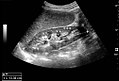
| + | [[File:Maximum length of spleen on ultrasonography.jpg|thumb|180px|Maximum length of the spleen.]] | ||
| + | *'''Spleen''', mainly for size, where 11 cm is a common cutoff. <br>''In case of suspected pathology, see: [[Ultrasonography of the spleen]]'' | ||
| + | Also, it is generally easy to have a quick glance at the kidneys just to exclude hydronephrosis. <br>''In case of suspected pathology, see: [[Ultrasonography of the urinary system]]'' | ||
| + | |||
| + | ;Report | ||
| + | {|class="wikitable" | ||
| + | | | ||
| + | Example in a normal case: | ||
| + | <br>Gallbladder with thin wall and no gallstones. Normal width of the intra-and extrahepatic bile ducts. | ||
| + | <br>Liver with normal echogenicity and no focal lesions. | ||
| + | <br>Normal head and body of the pancreas. | ||
| + | <br>Normally sized spleen. | ||
| + | {{Public Domain example}} | ||
| + | |} | ||
| + | {{Reporting}} | ||
| + | |||
| + | ===Lower abdomen=== | ||
| + | Ultrasonographies of the lower abdomen can generally be focused on the condition requested in the referral. | ||
| + | |||
| + | ==Diseases and conditions== | ||
| + | |||
| + | ===Biliary tract=== | ||
| + | *[[Ultrasonography of gallstones|Ultrasonography of '''gallstones''']] | ||
| + | *[[Ultrasonography of cholecystitis|Ultrasonography of '''cholecystitis''']] | ||
| + | |||
| + | ===Appendicitis=== | ||
| + | *[[Ultrasonography of appendicitis|Ultrasonography of '''appendicitis''']] | ||
| + | |||
| + | ===Aneurysm=== | ||
| + | *[[Ultrasonography of abdominal aneurysm|Ultrasonography of '''abdominal aneurysm''']] | ||
| + | |||
| + | ===Cirrhosis=== | ||
| + | *[[Ultrasonography of cirrhosis|Ultrasonography of '''cirrhosis''']] | ||
| + | |||
| + | ===Epigastric bulding=== | ||
| + | *[[Ultrasonography of epigastric bulging|Ultrasonography of '''epigastric bulging''']] | ||
| + | {{Bottom}} | ||
Latest revision as of 12:16, 13 September 2019
Author:
Mikael Häggström [notes 1]
Contents
Planning
- Choice of exam
For patients presenting with symptoms that are less specific for any certain organ, such as unspecific acute abdominal pain, consider an abdominal/pelvic CT instead.
- At least 6 hours of fasting[notes 2]
Exceptions:
- The target structures are known to be superficial to any intestines
- The case is emergent enough to justify a suboptimal exam
Locations


General screening
Upper abdomen
For symptoms of the upper abdomen, it is a proper custom to perform a general screening of the following locations:
- Pancreas, mainly for dilatation of the pancreatic duct, or obvious tumors.
- Liver, for echogenicity and focal changes.
In case of suspected pathology, see: Ultrasonography of the liver - Hepatorenal recess for ascites.
- Biliary tract, for gallstones (see Ultrasonography of gallstones) and dilatation of the intrahepatic or extrahepatic bile ducts. The common bile duct is normally up to 8 mm.[1] Further information: Basic screening of the biliary tract.

- Spleen, mainly for size, where 11 cm is a common cutoff.
In case of suspected pathology, see: Ultrasonography of the spleen
Also, it is generally easy to have a quick glance at the kidneys just to exclude hydronephrosis.
In case of suspected pathology, see: Ultrasonography of the urinary system
- Report
|
Example in a normal case:
|
- See also: General notes on reporting
Lower abdomen
Ultrasonographies of the lower abdomen can generally be focused on the condition requested in the referral.
Diseases and conditions
Biliary tract
Appendicitis
Aneurysm
Cirrhosis
Epigastric bulding
Notes
- ↑ For a full list of contributors, see article history. Creators of images are attributed at the image description pages, seen by clicking on the images. See Radlines:Authorship for details.
- ↑ Otherwise high risk of intestines obscuring the organ of interest.
- NU Hospital Group, Sweden
References
- ↑ Hoeffel, Christine; Azizi, Louisa; Lewin, Maité; Laurent, Valérie; Aubé, Christophe; Arrivé, Lionel; Tubiana, Jean-Michel (2006). "Normal and Pathologic Features of the Postoperative Biliary Tract at 3D MR Cholangiopancreatography and MR Imaging ". RadioGraphics 26 (6): 1603–1620. doi:. ISSN 0271-5333.