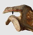
X-ray of shoulder impingement
Author:
Mikael Häggström [notes 1]
Planning
Indication
Impingement syndrome can be diagnosed by a targeted medical history and physical examination,[1][2] but it has also been argued that medical imaging, primarily projectional radiography ("X-ray") is a necessary part of the workup.[3]
Method
X-ray of the shoulder that should include:
- Frontal with and without external rotation of the shoulder
- Scapular outlet view
Evaluation
- Measurement of the subacromial space. This is normally 9–10 mm in shoulder radiographs.[4] It is significantly greater in men, with a slight reduction with age.[4] In middle age, a subacromial space less than 6 mm is pathological.[4]
- In case of decreased subacromial space, also measure the position of the humerus as described at X-ray of rotator cuff tear
- Shape of the acromion, at least for the presence of any acromial spur
Also look for common differential diagnoses or worsening factors of shoulder impingement:
- Osteoarthritis. For severity grading, see X-ray of osteoarthritis of the shoulder
- Presence of any calcifications in the supraspinatus tendon, which indicates a differential diagnosis of calcific tendonitis.
Report
- Either normal subacromial space, or a decrease thereof, with measurement of the space in the latter case.
- Presence or absence of an acromial spur.
A more comprehensive report also mentions:
- The acromial shape regardless[notes 2]
- Even the absence of osteoarthritis.
- See also: General notes on reporting
Notes
- ↑ For a full list of contributors, see article history. Creators of images are attributed at the image description pages, seen by clicking on the images. See Radlines:Authorship for details.
- ↑ 2.0 2.1 2.2 2.3 Generally, report the shape of the acromion in the form of type 1, 2 or 3 only if responding to an orthopedic clinic where this is the customary classification.
References
- ↑ Craig Hacking and Frank Gaillard (2019-03-06). Subacromial impingement. Radiopaedia.
- ↑ . Shoulder Impingement Syndrome. Stanford University Medical Center (2019-03-06).
- ↑ Garving, Christina; Jakob, Sascha; Bauer, Isabel; Nadjar, Rudolph; Brunner, Ulrich H. (2017). "Impingement Syndrome of the Shoulder ". Deutsches Aerzteblatt Online. doi:. ISSN 1866-0452.
- ↑ 4.0 4.1 4.2 Claes J. Petersson & lnga Redlund- Johnell (1984). "The subacromial space in normal shoulder radiographs ". Acta Orthop Scanda 55 (1): 57–8. PMID 6702430.


