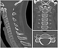
CT of the neck
Jump to navigation
Jump to search
Author:
Mikael Häggström [notes 1]
Contents
Normal anatomy
Diseases and presentations
Lymphadenopathy
| Generally (non-retropharyngeal) | 10 mm[1] – 20 mm[2][3] |
| Jugulodigastric lymph nodes | 11mm[1] or 15 mm[3] |
| Retropharyngeal | 8 mm[3]
|
Locations
Notes
- ↑ For a full list of contributors, see article history. Creators of images are attributed at the image description pages, seen by clicking on the images. See Radlines:Authorship for details.
References
- ↑ 1.0 1.1 1.2 "Current concepts in lymph node imaging ". Journal of Nuclear Medicine : Official Publication, Society of Nuclear Medicine 45 (9): 1509–18. September 2004. PMID 15347718.
- ↑ . Assessment of lymphadenopathy. BMJ Best Practice. Retrieved on 2017-03-04. Last updated: Last updated: Feb 16, 2017
- ↑ 3.0 3.1 3.2 Page 432 in: Luca Saba (2016). Image Principles, Neck, and the Brain . CRC Press. ISBN 9781482216202.


