MRI sequences
Jump to navigation
Jump to search
Author:
Mikael Häggström [notes 1]
Sequences
| Group | Sequence | Abbr. | Main clinical distinctions | Example |
|---|---|---|---|---|
| Spin echo | T1 weighted | T1 |
Standard foundation and comparison for other sequences |

|
| T2 weighted | T2 |
Standard foundation and comparison for other sequences |

| |
| Proton density weighted | PD | Joint disease and injury.[3]
|

| |
| Gradient echo | Steady-state free precession | SSFP | Creation of cardiac MRI videos (pictured).[5] | 
|
| Inversion recovery | Short tau inversion recovery | STIR | High signal in edema, such as in more severe stress fracture[6] Shin splints pictured: | 
|
| Fluid-attenuated inversion recovery | FLAIR | High signal in lacunar infarction, multiple sclerosis (MS) plaques, subarachnoid haemorrhage and meningitis (pictured).[7] | 
| |
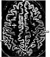
| Double inversion recovery | DIR | High signal of multiple sclerosis plaques (pictured)[8] | 
| |
| Diffusion weighted (DWI) | Conventional | DWI | High signal within minutes of cerebral infarction (pictured).[9] | 
|
| Apparent diffusion coefficient | ADC | Low signal minutes after cerebral infarction (pictured)[10] | 
| |
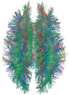
| Diffusion tensor | DTI | 
| ||
| Perfusion weighted (PWI) | Dynamic susceptibility contrast | DSC | In cerebral infarction, the infarcted core and the penumbra have decreased perfusion (pictured).[13] | 
|
| Dynamic contrast enhanced | DCE | |||
| Arterial spin labelling | ASL | |||
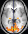
| Functional MRI (fMRI) | Blood-oxygen-level dependent imaging | BOLD | Localizing highly active brain areas before surgery[14] | 
|
| Magnetic resonance angiography (MRA) and venography | Time-of-flight | TOF | Detection of aneurysm, stenosis, or dissection[15] | 
|
| Phase-contrast magnetic resonance imaging | PC-MRA | Detection of aneurysm, stenosis and dissection (pictured)[15] |  Vastly undersampled Isotropic Projection Reconstruction (VIPR) | |
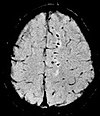
| Susceptibility-weighted | SWI | Detecting small amounts of hemorrhage (diffuse axonal injury pictured) or calcium[16] | 
| |
Notes
- ↑ For a full list of contributors, see article history. Creators of images are attributed at the image description pages, seen by clicking on the images. See Radlines:Authorship for details.
References
- ↑ 1.0 1.1 1.2 1.3 . Magnetic Resonance Imaging. University of Wisconsin. Retrieved on 2016-03-14.
- ↑ 2.0 2.1 2.2 2.3 Keith A. Johnson. Basic proton MR imaging. Tissue Signal Characteristics. Harvard Medical School. Archived from the original on 2016-03-05. Retrieved on 2016-03-14.
- ↑ Jeremy Jones and Prof Frank Gaillard et al.. MRI sequences (overview). Radiopaedia. Retrieved on 2017-01-13.
- ↑ Lefevre, Nicolas; Naouri, Jean Francois; Herman, Serge; Gerometta, Antoine; Klouche, Shahnaz; Bohu, Yoann (2016). "A Current Review of the Meniscus Imaging: Proposition of a Useful Tool for Its Radiologic Analysis ". Radiology Research and Practice 2016: 1–25. doi:. ISSN 2090-1941.
- ↑ Dr Tim Luijkx and Dr Yuranga Weerakkody. Steady-state free precession MRI. Radiopaedia. Retrieved on 2017-10-13.
- ↑ Ferco Berger, Milko de Jonge, Robin Smithuis and Mario Maas. Stress fractures. Radiopaedia. Retrieved on 2017-10-13.
- ↑ . Fluid attenuation inversion recoveryg. radiopaedia.org. Retrieved on 2015-12-03.
- ↑ Dr Bruno Di Muzio and Dr Ahmed Abd Rabou. Double inversion recovery sequence. Radiopaedia. Retrieved on 2017-10-13.
- ↑ Dr Yuranga Weerakkody and Prof Frank Gaillard et al.. Ischaemic stroke. Radiopaedia. Retrieved on 2017-10-15.
- ↑ An, H.; Ford, A. L.; Vo, K.; Powers, W. J.; Lee, J.-M.; Lin, W. (2011). "Signal Evolution and Infarction Risk for Apparent Diffusion Coefficient Lesions in Acute Ischemic Stroke Are Both Time- and Perfusion-Dependent ". Stroke 42 (5): 1276–1281. doi:. ISSN 0039-2499.
- ↑ Derek Smith and Dr Usman Bashir. Diffusion tensor imaging. Radiopaedia. Retrieved on 2017-10-13.
- ↑ Chua, Terence C; Wen, Wei; Slavin, Melissa J; Sachdev, Perminder S (2008). "Diffusion tensor imaging in mild cognitive impairment and Alzheimerʼs disease: a review ". Current Opinion in Neurology 21 (1): 83–92. doi:. ISSN 1350-7540.
- ↑ Chen, Feng (2012). "Magnetic resonance diffusion-perfusion mismatch in acute ischemic stroke: An update ". World Journal of Radiology 4 (3): 63. doi:. ISSN 1949-8470.
- ↑ Dr Tim Luijkx and Prof Frank Gaillard. Functional MRI. Radiopaedia. Retrieved on 2017-10-16.
- ↑ 15.0 15.1 . Magnetic Resonance Angiography (MRA). Johns Hopkins Hospital. Retrieved on 2017-10-15.
- ↑ Dr Bruno Di Muzio and A.Prof Frank Gaillard. Susceptibility weighted imaging. Retrieved on 2017-10-15.