X-ray of the thorax
Author:
Mikael Häggström [notes 1]
Contents
Planning
Choice of investigation
- X-ray of the thorax is generally the initial investigation of choice in dyspnea.
- CT of the thorax is generally the investigation of choice in pulmonary embolism and aortic dissection. It is recommended as complementary to X-ray in chest trauma.[1]
Evaluation
Quality checking
The frontal X-ray should be straight enough to avoid excessive coverage of the lung fields by the mediastinum.
The diaphragm should be intersected by the 5th to 7th anterior ribs in the mid-clavicular line.
Magnification
In projectional radiography ("X-ray") of the thorax (chest), the estimated geometric magnification factor is generally between 1.05 and 1.40,[2] meaning that measurements made in the image will be this much larger than the real distance in the patient. Therefore, compare to other structures (such as pleural fluid reaching half way to hilar level) where possible rather than giving numbers.
Normal anatomy
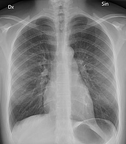
A normal posteroanterior (PA) chest radiograph.

Normal lateral chest radiograph.

Projectionally rendered CT, showing the transition of thoracic structures between the anteroposterior and lateral view.
_chest_radiograph_(X-ray).jpg)
.jpg)
.gif)
Basic screening
One method uses an alphabetic order:
- Airways: Any shift to the side?
- Bones: Check for obvious fractures

where:[3]
MRD = greatest perpendicular diameter from midline to right heart border
MLD = greatest perpendicular diameter from midline to left heart border
ID = internal diameter of chest at level of right hemidiaphragm
- Cardiac: Check both cardiothoracic ratio and any dilation of blood vessels.
- Diaphragm: Check for any pleural fluid
- Edges: Check for pneumothorax and, if not already evaluated under C, also Kerley B lines.

Subtle pneumothorax, only determined by absence of vascularity in upper left lung region.

Kerley B lines


- Fields: Any lung consolidations
- Gastric: Whether there is any apparent hiatal hernia

Hiatal hernia.

- Hili: Prominence, such as by lymphadenopathy
- In lateral image: as a reminder to check the lateral projection, if available, mainly in the lung fields, for any apparent pathology
Report
- Even absence of lung opacities and pleural fluid.
- Upright images: Normal or increased heart size, and generally even absence of congestion.
- Supine: Report suspected cardiomegaly and/or dilated vessels only if the referral specifically requests it. Even then, note that it is difficult to determine with patient lying down.
- See also: General notes on reporting
Diseases and targets
If specifically suspected or requested in the referral:
- X-ray of the thorax in sarcoidosis
- X-ray of the thorax in tuberculosis
- X-ray of cardiac pacemakers
- X-ray of central venous catheters
Regions
- Ribs
- Sternoclavicular joint
Notes
- ↑ For a full list of contributors, see article history. Creators of images are attributed at the image description pages, seen by clicking on the images. See Radlines:Authorship for details.
References
- ↑ . Blunt Chest Trauma, ACR Appropriateness Criteria. American College of Radiology. Date of origin: 2013
- ↑ M Sandborg, D R Dance, and G Alm Carlsson. Implementation of unsharpness and noise into the model of the imaging system: Applications to chest and lumbar spine screen-film radiography. Faculty of Health Sciences, Linköping University. Report 90. Jan. 1999. ISRN: LIU-RAD-R-090
- ↑ . Chest Measurements. Oregon Health & Science University. Retrieved on 2017-01-13.