Template:Grading of hydronephrosis
Jump to navigation
Jump to search
Author:
Mikael Häggström [notes 1]
Hydronephrosis grading
_grading_of_hydronephrosis.jpg)
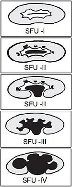
The Society of Fetal Ultrasound has developed a grading system for hydronephrosis, initially intended for use in neonatal and infant hydronephrosis, but it is now used for grading hydronephrosis in adults as well:[1]
- Grade 0 – No renal pelvis dilation. Cutoff values for different patient populations are:
- Fetuses: An anteroposterior diameter of less than 4 mm in fetuses up to 32 weeks of gestational age and 7 mm afterwards.[2]
- Adults, defineddifferently by different sources, with anteroposterior diameters ranging between 10 and 20 mm.[3] About 13% of normal healthy adults have a transverse pelvic diameter of over 10 mm.[4]
- Pregnant women in the last two trimesters: The maximum normal expected renal pelvic diameter (97.5 percent prediction interval) is 27 mm on the right and 18 mm on the left.[5]
- Grade 1 (mild) – Mild renal pelvis dilation (anteroposterior diameter less than 10 mm in fetuses[2]) without dilation of the calyces nor parenchymal atrophy
- Grade 2 (mild) – Moderate renal pelvis dilation (between 10 and 15 mm in fetuses[2]), including a few calyces
- Grade 3 (moderate) – Renal pelvis dilation with all calyces uniformly dilated. Normal renal parenchyma
- Grade 4 (severe) – As grade 3 but with thinning of the renal parenchyma
In Swedish practice,[notes 2] the most important is a subjective classification into mild, moderate or severe, with optional mention of numerical grade (unless specifically requested in the referral).
Notes
- ↑ For a full list of contributors, see article history. Creators of images are attributed at the image description pages, seen by clicking on the images. See Radlines:Authorship for details.
- ↑ NU Hospital Group, Sweden
References
- ↑ Laurence S Baskin. Overview of fetal hydronephrosis. Version Version 29.0. UpToDate. Retrieved on 2017-04-25. Last updated Apr 20, 2017
- ↑ 2.0 2.1 2.2 Page 189 in: V. D'Addario (2014). Donald School Basic Textbook of Ultrasound in Obstetrics & Gynecology . JP Medical Ltd. ISBN 9789351523376.
- ↑ Page 78 in: Justin Bowra, Russell E (2011). Emergency Ultrasound Made Easy, Edition 2 . Elsevier Health Sciences. ISBN 9780702048722.
- ↑ "Sonographic evaluation of renal appearance in 665 adult volunteers. Correlation with age and obesity ". Acta Radiol 34 (5): 482–5. 1993. doi:. PMID 8369185.
- ↑ Erickson, L. M.; Nicholson, S. F.; Lewall, D. B.; Frischke, Lauraline (1979). "Ultrasound evaluation of hydronephrosis of pregnancy ". Journal of Clinical Ultrasound 7 (2): 128–132. doi:. ISSN 00912751.