X-ray of Hansson pins
Author:
Mikael Häggström [notes 1]
Hansson pins are also called LIH pins after its developer Lars Ingvar Hansson, and are widely used in Sweden and the UK.[1]
Contents
Planning
Frontal and lateral projections.
Post-operative evaluation

Pin positions
Pin position evaluation is generally not mandatory for radiologists unless specifically requested by the orthopedic surgeon.
The pins should be at least 2 cm apart on frontal projection, and have <10° angle compared to each other.[2] The inferior pin should be inserted at the level of the minor trochanter (insertion below it increases the risk of subsequent subtrochanteric fracture).[2]
For proper support, both pins should have support by the subchondral plate and the lateral cortex. In addition, the middle of the inferior pin should be supported by the medial cortex, and the middle of the posterior pin should have support from the medial cortex, with less than <5 mm distance from subchondral bone.[2]
Fracture alignment
"No misalignment" means:[2]
- Dislocation of less than 2 mm
- 0-15° valgus angle on frontal projection
- Dorsal tilt of <10° on lateral projection
Leg length discrepancy
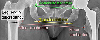
Leg length discrepancy after Hansson pins is calculated similarly to in X-ray of hip prostheses (pictured), calculated as the vertical distance between the middle of the minor trochanters, using the acetabular tear drops[3] or the transischial line[4] as references for the horizontal plane. A discrepancy of up to 1 cm is generally tolerated.[3][4]

Report
Example:
Inserted Hansson pins (and if evaluated, they) have proper positions.
No fracture misalignment or significant leg length discrepancy.
Notes
- ↑ For a full list of contributors, see article history. Creators of images are attributed at the image description pages, seen by clicking on the images. See Radlines:Authorship for details.
References
- ↑ "Lars Ingvar Hansson 1937–1987 ". Acta Orthopaedica Scandinavica 58 (6): 685–692. 2009. doi:. ISSN 0001-6470.
- ↑ 2.0 2.1 2.2 2.3 Marsel Toplić, Carl Mellner. LIH-spikning (Hansson Pin). ortobas.se.
- ↑ 3.0 3.1 Vanrusselt, Jan; Vansevenant, Milan; Vanderschueren, Geert; Vanhoenacker, Filip (2015). "Postoperative radiograph of the hip arthroplasty: what the radiologist should know ". Insights into Imaging 6 (6): 591–600. doi:. ISSN 1869-4101. PMID 26487647.
- ↑ 4.0 4.1 Iain Watt, Susanne Boldrik, Evert van Langelaan and Robin Smithuis. Hip - Arthroplasty -Normal and abnormal imaging findings. Radiology Assistant. Retrieved on 2017-05-21.