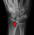
X-ray of scapholunate instability
Jump to navigation
Jump to search
Author:
Mikael Häggström [notes 1]
Contents
Planning
Required projections are posterior anterior (PA), a lateral view X-ray, and then either fist view X-ray[1] or X-rays in ulnar and radial deviation.
Evaluation
Measure the scapholunate distance. A distance of more than 3 mm, which is called scapholunate dissociation.[2] A static instability is generally readily visible, but a dynamic scapholunate instability can only be seen radiographically in certain wrist positions or under certain loading conditions, such as when clenching the wrist, or loading the wrist in ulnar deviation.[3]
Report
- Presence or absence of scapholunate dissociation. If present, specify if it is static, or in which positions it appears.
- See also: General notes on reporting
Notes
- ↑ For a full list of contributors, see article history. Creators of images are attributed at the image description pages, seen by clicking on the images. See Radlines:Authorship for details.
References
- ↑ Novelline, RA (2004). Squire's fundamentals of radiology, 6th Edition (6th ed.). United States of America: President and fellows of Harvard college. ISBN 0-674-01279-8.
- ↑ Owen Kang and Henry Knipe et al.. Scapholunate advanced collapse. Radiopaedia. Retrieved on 2018-01-05.
- ↑ Fairplay, Tracy; Cozzolino, Roberto; Atzei, Andrea; Luchetti, Riccardo (2013). "Current Role of Open Reconstruction of the Scapholunate Ligament ". Journal of Wrist Surgery 02 (02): 116–125. doi:. ISSN 2163-3916.


