X-ray of thumb fractures
Author:
Mikael Häggström [notes 1]
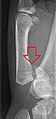
Projectional radiography ("X-ray") of fracture of the thumb:
Contents
Detection
Even if there's an initial obvious fracture, evaluate:
- Bone contours for disruptions
- Bone areas for unusual lines that are either hypoattenuating (in case of separation) or hyperattenuating (in case of compression)
In children
- Main article: X-ray of fractures in children
In patients with remaining growth plates, look for fracture involvement thereof (if present, see X-ray of fractures in children).
Classification
Metacarpal
- Intra-articular fractures
Bennet's fracture: A fracture of the base of the first metacarpal bone which is intra-articular, extending into the carpometacarpal (CMC) joint.[1] It is the most common type of fracture of the thumb, and is nearly always accompanied by some degree of subluxation or frank dislocation of the carpometacarpal joint.

Rolando fracture: A comminuted, Intra-articular fracture fracture through the base of the first metacarpal bone.[2])


- Extra-articular fractures

Avulsion fracture at the insertion of the ulnar collateral ligament

- Epibasal thumb fracture, or pseudo-Bennett fracture: Fracture of the proximal part of the first metacarpal, where it is important to exclude intra-articular involvement.[3]
Misalignment
Types of fracture misalignment:[4]
|
Reporting
Notes
- ↑ For a full list of contributors, see article history. Creators of images are attributed at the image description pages, seen by clicking on the images. See Radlines:Authorship for details.
References
- ↑ . Bennett fracture-subluxation. GPnotebook.
- ↑ http://www.wheelessonline.com/ortho/rolandos_fracture
- ↑ Tim Luijkx and A.Prof Frank Gaillard et al.. Epibasal fracture of the thumb. Retrieved on 2018-07-09.
- ↑ . Introduction to Trauma X-ray. Radiology Masterclass. Retrieved on 2018-07-03.