Difference between revisions of "X-ray of hip pain"
(+Findings) |
m (Spaced) |
||
| (7 intermediate revisions by the same user not shown) | |||
| Line 3: | Line 3: | ||
|author2= | |author2= | ||
}} | }} | ||
| + | |||
This article focuses on long-term hip-pain rather than fractures (see [[X-ray of fractures]]). | This article focuses on long-term hip-pain rather than fractures (see [[X-ray of fractures]]). | ||
| Line 10: | Line 11: | ||
It is performed as an [[X-ray of the hip joint]]. | It is performed as an [[X-ray of the hip joint]]. | ||
| − | ==Evaluation | + | ==Evaluation== |
The main focus is on the joint, but also includes an overview of the projected pelvic bones and femur. | The main focus is on the joint, but also includes an overview of the projected pelvic bones and femur. | ||
| − | ===Potential findings | + | ===Potential findings in adults=== |
<gallery mode=packed heights=180> | <gallery mode=packed heights=180> | ||
File:X-ray of a normal hip joint.jpg|Normal hip | File:X-ray of a normal hip joint.jpg|Normal hip | ||
| − | File:X-ray of pincer impingement.jpg|X-ray in pincer impingement type of hip dysplasia, showing a pincer at the lateral part of the acetabulum | + | File:X-ray of pincer impingement.jpg|X-ray in pincer impingement type of hip dysplasia, showing a pincer at the lateral part of the acetabulum<ref name="Ruiz SantiagoSantiago Chinchilla2016"/> |
| − | File:X-ray of cam.jpg|X-ray of cam, with bulging at the lateral part of the femur | + | File:X-ray of cam.jpg|X-ray of cam, with bulging at the lateral part of the neck of the femur<ref name="Ruiz SantiagoSantiago Chinchilla2016"/> |
| − | File:X-ray of hip osteoarthritis.jpg|Hip in osteoarthritis with joint space narrowing and sclerosis. See '''[[X-ray of osteoarthritis of the hip joint]]''' | + | File:X-ray of hip osteoarthritis.jpg|Hip in osteoarthritis with joint space narrowing and sclerosis. <br>See '''[[X-ray of osteoarthritis of the hip joint]]''' |
| − | File:X-ray of septic arthritis of the hip.jpg|Septic arthritis, similar to osteoarthritis but occurs faster and is generally more erosive. | + | File:X-ray of septic arthritis of the hip.jpg|Septic arthritis, similar to osteoarthritis but occurs faster and is generally more erosive.<ref name="Ruiz SantiagoSantiago Chinchilla2016"/> |
File:X-ray of synovial chondromatosis.jpg|X-ray of synovial chondromatosis, with a calcified cartilaginous chondroma inferiorly.<ref name="Ruiz SantiagoSantiago Chinchilla2016"/> | File:X-ray of synovial chondromatosis.jpg|X-ray of synovial chondromatosis, with a calcified cartilaginous chondroma inferiorly.<ref name="Ruiz SantiagoSantiago Chinchilla2016"/> | ||
| + | </gallery> | ||
| + | |||
| + | ==Potential findings in children== | ||
| + | <gallery mode=packed heights=180> | ||
| + | File:Roe-perthes.jpg|Legg–Calvé–Perthes disease occurs mainly between the ages of 4 and 10 years, with osteonecrosis of the left head of the femur<ref name="Ruiz SantiagoSantiago Chinchilla2016"/><br>See [[X-ray of Legg–Calvé–Perthes disease|X-ray of '''Legg–Calvé–Perthes disease''']] | ||
| + | File:Slipped capital femoral epiphysis.jpg|Slipped capital femoral epiphysis usually affect 11- to 14-year-old patients.<ref name="Ruiz SantiagoSantiago Chinchilla2016"/> | ||
| + | File:X-ray of left hip effusion secondary to trauma.jpg|Left hip effusion, showing widening of the medial joint space.<ref name="Ruiz SantiagoSantiago Chinchilla2016"/> | ||
| + | File:X-ray of measurements in hip dysplasia.jpg|link=X-ray of hip dysplasia|Hip dysplasia, with insufficient acetabular coverage of the head of the femur. <br>See [[X-ray of hip dysplasia|X-ray of '''hip dysplasia''']] | ||
</gallery> | </gallery> | ||
{{Bottom}} | {{Bottom}} | ||
Latest revision as of 13:48, 18 December 2018
Author:
Mikael Häggström [notes 1]
This article focuses on long-term hip-pain rather than fractures (see X-ray of fractures).
Contents
Planning
Choice of investigation
Projectional radiography ("X-ray") of hip pain is the first imaging technique of choice, not only in older people with suspected osteoarthritis but also in young people without any such suspicion. In this case plain radiography allows categorization of normal anatomy versus hip dysplasia, or with impingement signs, pincer, cam, or a combination of both.[1]
Also consider the possibility of inguinal hernia.
It is performed as an X-ray of the hip joint.
Evaluation
The main focus is on the joint, but also includes an overview of the projected pelvic bones and femur.
Potential findings in adults

Normal hip

X-ray in pincer impingement type of hip dysplasia, showing a pincer at the lateral part of the acetabulum[1]

X-ray of cam, with bulging at the lateral part of the neck of the femur[1]

Hip in osteoarthritis with joint space narrowing and sclerosis.
See X-ray of osteoarthritis of the hip joint
Septic arthritis, similar to osteoarthritis but occurs faster and is generally more erosive.[1]
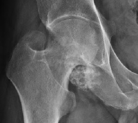
X-ray of synovial chondromatosis, with a calcified cartilaginous chondroma inferiorly.[1]


Potential findings in children

Legg–Calvé–Perthes disease occurs mainly between the ages of 4 and 10 years, with osteonecrosis of the left head of the femur[1]
See X-ray of Legg–Calvé–Perthes disease
Slipped capital femoral epiphysis usually affect 11- to 14-year-old patients.[1]
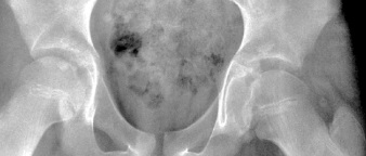
Left hip effusion, showing widening of the medial joint space.[1]

Hip dysplasia, with insufficient acetabular coverage of the head of the femur.
See X-ray of hip dysplasia


Notes
- ↑ For a full list of contributors, see article history. Creators of images are attributed at the image description pages, seen by clicking on the images. See Radlines:Authorship for details.
References
- ↑ 1.0 1.1 1.2 1.3 1.4 1.5 1.6 1.7 Initially largely copied from: Ruiz Santiago, Fernando; Santiago Chinchilla, Alicia; Ansari, Afshin; Guzmán Álvarez, Luis; Castellano García, Maria del Mar; Martínez Martínez, Alberto; Tercedor Sánchez, Juan (2016). "Imaging of Hip Pain: From Radiography to Cross-Sectional Imaging Techniques ". Radiology Research and Practice 2016: 1–15. doi:. ISSN 2090-1941. Attribution 4.0 International (CC BY 4.0) license
