Difference between revisions of "CT of the abdomen and pelvis"
Jump to navigation
Jump to search


(Linked) |
(expanded) |
||
| Line 3: | Line 3: | ||
|author2= | |author2= | ||
}} | }} | ||
| − | |||
| − | |||
| − | |||
| − | == | + | ==Locations== |
| − | {{ | + | {{Abdomen and pelvis locations}} |
| + | |||
| + | ==Basic screening== | ||
| + | [[Compare]] with any previous exam. | ||
| + | |||
| + | Example approach: | ||
| + | *'''Liver, spleen and adrenals''': Scan through the entire volumes and look mainly for focal changes. | ||
| + | :*The thickness of the adrenal glands is normally up to 1.0 cm.<ref>{{cite journal|url=http://www.medscape.com/viewarticle/543510_2|title=CT and MRI of Adrenal Masses|author=Antonio Carlos A. Westphalen and Bonnie N. Joe|journal=Appl Radiol|year=2006|volume=35|issue=8|pages=10–26}}</ref> | ||
| + | Further information: | ||
| + | <gallery> | ||
| + | File:CT of the liver.jpg|link=CT of the liver|[[CT of the liver|CT of the '''liver''']] | ||
| + | File:CT of adrenal adenoma.jpg|link=CT of the adrenals|[[CT of the '''adrenals''']] | ||
| + | </gallery> | ||
==Large intestine== | ==Large intestine== | ||
| Line 15: | Line 24: | ||
==Small intestine== | ==Small intestine== | ||
On CT scans, a small intestinal diameter of over 2.5 cm is considered abnormally dilated.<ref name=medscape>{{cite web|url=http://emedicine.medscape.com/article/374962-overview|title=Small-Bowel Obstruction Imaging|website=Medscape|date=2016-09-22|author=Ali Nawaz Khan|accessdate=2017-02-07}}</ref><ref>{{cite web|url=http://www.radiologymasterclass.co.uk/tutorials/abdo/abdomen_x-ray_abnormalities/pathology_small_bowel_obstruction|title=Abdominal X-ray - Abnormal bowel gas pattern|website=radiologymasterclass.co.uk|accessdate=2017-02-07}}</ref><ref name="GazelleGoldberg1994">{{cite journal|last1=Gazelle|first1=G S|last2=Goldberg|first2=M A|last3=Wittenberg|first3=J|last4=Halpern|first4=E F|last5=Pinkney|first5=L|last6=Mueller|first6=P R|title=Efficacy of CT in distinguishing small-bowel obstruction from other causes of small-bowel dilatation.|journal=American Journal of Roentgenology|volume=162|issue=1|year=1994|pages=43–47|issn=0361-803X|doi=10.2214/ajr.162.1.8273687}}</ref> | On CT scans, a small intestinal diameter of over 2.5 cm is considered abnormally dilated.<ref name=medscape>{{cite web|url=http://emedicine.medscape.com/article/374962-overview|title=Small-Bowel Obstruction Imaging|website=Medscape|date=2016-09-22|author=Ali Nawaz Khan|accessdate=2017-02-07}}</ref><ref>{{cite web|url=http://www.radiologymasterclass.co.uk/tutorials/abdo/abdomen_x-ray_abnormalities/pathology_small_bowel_obstruction|title=Abdominal X-ray - Abnormal bowel gas pattern|website=radiologymasterclass.co.uk|accessdate=2017-02-07}}</ref><ref name="GazelleGoldberg1994">{{cite journal|last1=Gazelle|first1=G S|last2=Goldberg|first2=M A|last3=Wittenberg|first3=J|last4=Halpern|first4=E F|last5=Pinkney|first5=L|last6=Mueller|first6=P R|title=Efficacy of CT in distinguishing small-bowel obstruction from other causes of small-bowel dilatation.|journal=American Journal of Roentgenology|volume=162|issue=1|year=1994|pages=43–47|issn=0361-803X|doi=10.2214/ajr.162.1.8273687}}</ref> | ||
| + | |||
| + | ===Report=== | ||
==Aneurysm== | ==Aneurysm== | ||
Revision as of 12:16, 21 December 2018
Author:
Mikael Häggström [notes 1]
Contents
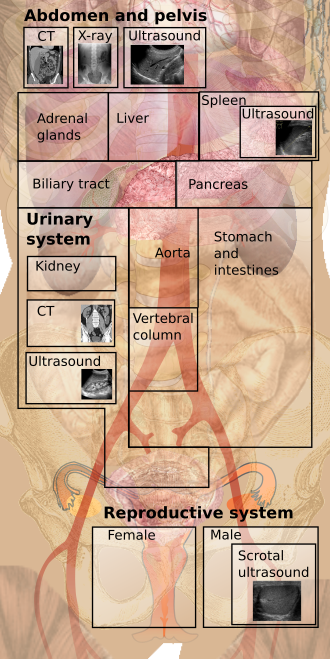
Locations

Basic screening
Compare with any previous exam.
Example approach:
- Liver, spleen and adrenals: Scan through the entire volumes and look mainly for focal changes.
- The thickness of the adrenal glands is normally up to 1.0 cm.[1]
Further information:


Large intestine

Average inner diameters and ranges of different sections of the large intestine.[2]
Small intestine
On CT scans, a small intestinal diameter of over 2.5 cm is considered abnormally dilated.[3][4][5]
Report
Aneurysm
Notes
- ↑ For a full list of contributors, see article history. Creators of images are attributed at the image description pages, seen by clicking on the images. See Radlines:Authorship for details.
References
- ↑ Antonio Carlos A. Westphalen and Bonnie N. Joe (2006). "CT and MRI of Adrenal Masses ". Appl Radiol 35 (8): 10–26. Archived from the original. .
- ↑ "Deficient Pms2, ERCC1, Ku86, CcOI in field defects during progression to colon cancer ". J Vis Exp (41). 2010. doi:. PMID 20689513.
- ↑ Ali Nawaz Khan (2016-09-22). Small-Bowel Obstruction Imaging. Medscape. Retrieved on 2017-02-07.
- ↑ . Abdominal X-ray - Abnormal bowel gas pattern. radiologymasterclass.co.uk. Retrieved on 2017-02-07.
- ↑ Gazelle, G S; Goldberg, M A; Wittenberg, J; Halpern, E F; Pinkney, L; Mueller, P R (1994). "Efficacy of CT in distinguishing small-bowel obstruction from other causes of small-bowel dilatation. ". American Journal of Roentgenology 162 (1): 43–47. doi:. ISSN 0361-803X.
